


Continuous evaluations of athletes, including strength testing, can help control performance improvement or facilitate the restoration of normality after an injury. The aim of the present study was to prospectively determine the peak torque (PT), angle at which PT is achieved, and functional ratios of flexors and extensors thigh muscles during one season.
Material and methodsThirty semi-professional male athletes competing in long jumping (n = 10), javelin throwing (n = 10), and sprinting (n = 10) participated. PT was evaluated in relation to limb length; the angle at which PT was achieved was obtained from the force-curve displayed in the isokinetic dynamometer; functional ratios were calculated by dividing concentric hamstring strength by eccentric quadriceps strength (flexor ratio) or vice-versa for the extensor ratio. Assessment was performed at 60º/s and 300º/s.
ResultsSignificant variations were seen for both extensor and flexor PTs at different stages of the season, with moderate to large effect sizes observed (effect size (d) = 0.49–0.93). Functional ratios and the angle at which peak torque was achieved remained stable throughout the season.
ConclusionsThigh muscle strength is unstable throughout a track and field season, coaches or medical staff should consider these findings when programming training sessions or rehabilitating an athlete.
Muscle injuries are common in track and field, affecting mainly the thigh region.1 High speed running requires explosive hamstring (HAMS) and quadriceps femoris (QF) contractions, which poses a risk of strain to these muscle groups.2,3 For knee flexors, the late swing phase of the gait cycle (where the hip is flexed and the knee is in almost complete extension) demands high eccentric actions while working in a lengthened state. These demands increase as the runner accelerates, contributing to the high incidence of HAMS injuries during sprinting.4 By contrast, QF strains occur during the middle of the sprint cycle, when the hip is extended and the QF has to produce high eccentric force to brake the limb.5 These latest actions are common in the disciplines of sprinting, long jumping or javelin throwing. A recent review has confirmed that HAMS injury incidence is high in field-based sports, with 0.81 injuries every 1000 h of exposure.6 In addition, the time taken before the athlete can return to full training may be anything from three weeks to several months, depending on the specific location of the injury.7
Many factors render the athlete more susceptible to thigh muscle injuries. There is consistent evidence that a previous HAMS or QF injury may increase the risk of a subsequent QF injury or hamstring strain injury (HSI).8,9 Other include type II fibre predominance,10 performing at a competitive level,5,9 and deficits in eccentric strength.11
Isokinetic dynamometry, which provides objective measures for thigh muscle strength assessments, is widely used in sporting and research settings. It is considered to be the gold standard because it is convenient and reliable.12 This test allows the visualisation of torque production at each moment of the range of motion (ROM).13 Thus, HAMS/QF and QF/HAMS ratios can be calculated, which may be more relevant in sporting contexts.13 Since HAMS and QF strength play a significant role in decisions concerning the resumption of training or competitive activity after injury,14 it is important that information relating to healthy athletes is reported; clinicians can then access normative values and compare their results with those of uninjured athletes.
To the best of our knowledge, no previous studies have evaluated the evolution of torque and functional ratios throughout a sports season in track and field athletes. In a longitudinal setting, it is possible to hypothesise that these values are likely to change at different phases of the season. Coaches or medical staff could benefit from these observations both from a performance and injury prevention perspective. Therefore, the primary purpose of the present study was to determine the peak torque, the knee flexion angle at which peak torque was achieved, and the functional ratios of the thigh muscles, in three athletic specialities (sprinters, long jumpers, and javelin throwers) at three different stages of the regular season.
Materials and methodsSettingAthletes affiliated to the local university (University of Oviedo) were invited to participate. An information session was performed before data collection started, with a detailed explanation of the evaluation procedures. Attendants could ask any question related to the study development, and participation was voluntary. The corresponding Institutional Review Board approved the design and development of this study (Comité de Ética de la Investigación del Principado de Asturias: 2021.543). All participants signed the informed consent prior to the start of the first assessment day. The Principles of the Declaration of Helsinki were followed at all times.
SubjectsThirty semi-professional male athletes (sprinters, long jumpers, and javelin throwers) agreed to participate in the study. All participants were on scholarships and competing at national and international level. Only athletes who were available for strength testing during the study period were included. The exclusion criteria were to have sustained a thigh injury in the three months before the start of data collection or to suffer any lower limb injury during the study duration. After applying these criteria, all 30 participants completed the three measurement points, and their data were included in the study (Table 1).
Participants anthropometric characteristics.
Information is reported as mean (standard deviation).
Isokinetic evaluations of knee extensor and flexor muscles were tested at three timepoints during the season (1st: Last week of preseason; 2nd: Second week of competition; 3rd: End of season). Subjects were tested using a Kin-Com dynamometer (Chattanooga Group, Inc.) After a detailed explanation of the test and a general warm-up period, subjects were seated with their hips fixed at 87º of flexion. Participants’ trunks, hips, and thighs were firmly secured with Velcro straps (VELCRO USA, Inc., Manchester, New Hampshire) to the seat.
The axis of the dynamometer was aligned with the axis of the knee joint. The lower limb was strapped to the lever arm of the dynamometer at the level of 65% of the leg length, measured from the lateral femoral condyle to the lateral malleolus. Gravitational corrections were made for the effect of limb weight on torque measurements and a pre-load equivalent to the lower limb weight plus 10% was established. Subjects were given visual feedback during testing to increase strength output.
Both flexor and extensor knee muscles were evaluated at slow (60º/s) and fast (300º/s) velocities during concentric (CON) and eccentric (ECC) actions. We decided to measure strength at slow speed first, followed by fast velocity evaluations. Knee flexors and CON contractions were recorded first. The testing protocol consisted of three submaximal concentric and eccentric contractions with a 15-second recovery period between contractions, followed by three maximal concentric and eccentric contractions with a 30-second rest interval between each contraction. The maximal force curve was accepted and the knee motion ranged from 5º to 85º (with 0º indicating full extension).
Body fat percentage was calculated measuring six skinfolds (Triceps, subscapular, iliac crest, abdominal, front thigh, and medial calf) on the right side of the participants' bodies using the Yuhasz equation: % of body fat = (¿ of six skinfolds × 0.1051) + 2.585.15 An expert assessor performed the evaluations (BM), this method has shown to be valid compared to the gold standard of dual energy X-ray absorptiometry.16
Outcome measuresPeak torque (PT), the knee flexion angle at which PT was produced, and functional ratios were extracted from the isokinetic dynamometer for analysis. PT was defined as the maximum force that a muscle can generate around a joint range of movement and is expressed in Newtons (N), angle at which PT was produced is defined as the specific joint angle at which the muscles produce the highest torque during the joint range of movement and is expressed in degrees (º).
The functional ratio representative for knee flexion (FL) was defined as the ratio of CON HAMS force in relation to ECC QF force. By contrast, knee extension (EX) was defined as the ratio of CON quadriceps force in relation to ECC hamstring force. Both ratios were calculated at 60º/s and 300º/s.
Statistical analysisData was analysed using SPSS Statistics 26 (IBM). Significance level was set to p < 0.05. Normality assessment was tested with Kolmogorov-Smirnov test and by visual inspection of histograms. Demographic data is presented as mean ± standard deviation. Continuous variables were compared between athletic disciplines using repeated measures ANOVA, with post hoc analysis when significance was achieved for intergroup and intragroup comparisons. Effect sizes were calculated by use of Cohen's d and interpreted as small (d = 0.2), moderate (d = 0.5), and large (d = 0.8).
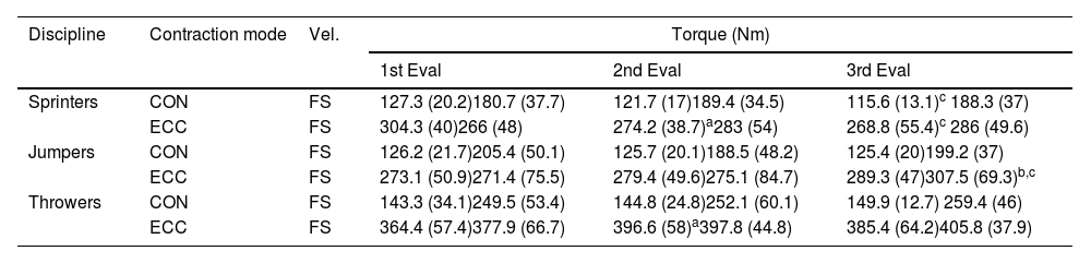
ResultsKnee flexor peak torqueThe PT of the knee flexors for each discipline can be seen in Table 2. Post hoc analyses revealed significant Group × Time interactions for both sprinters and javelin throwers. The sprinters’ CON fast knee flexor PT decreased significantly from the first to the second evaluation (d = 0.9), ECC fast knee flexor PT decreased significantly from the 1st to 2nd evaluations (d = 0.69) and decreased further until the third time-point assessment (d = 0.93). ECC slow decreased significantly between the 1st and 3rd evaluation points (d = 0.57). In javelin throwers, ECC strength significantly increased by the 2nd time-point at both fast (d = 0.78) and slow (d = 0.75) velocity.
Knee flexors peak torque throughout the season for the three different athletics disciplines.
Vel. = Velocity, Eval = Evaluation, CON = Concentric, ECC = Eccentric, F = Fast (300º/s), S = Slow (60º/s); Torque is displayed in Newtons × metre (Nm).
ap< 0.05 between 1st vs 2nd evaluation; bp< 0.05 between 2nd vs 3rd evaluation; cp< 0.05 between 1st vs 3rd evaluation.
Knee extensor PT is shown in Table 3. Post hoc comparisons revealed Group × Time interactions in all three disciplines. In sprinters, CON fast torque significantly decreased throughout the season (d = 0.68), and the same interaction was observed for ECC fast PT (d = 0.73). By contrast, the long jumpers’ ECC slow PT increased throughout the season (d = 0.49). Finally, in the javelin thrower group, ECC fast torque increased in the midseason (d = 0.56) but dropped during the final stage.
Knee extensors peak torque throughout the season for the three different athletics populations.
Vel. = Velocity, Eval = Evaluation, CON = Concentric, ECC = Eccentric, F = Fast (300º/s), S = Slow (60º/s); Torque is displayed in Newtons × metre (Nm).
ap< 0.05 1st vs 2nd evaluation; bp< 0.05 2nd vs 3rd evaluation; cp< 0.05 1st vs 3rd evaluation.
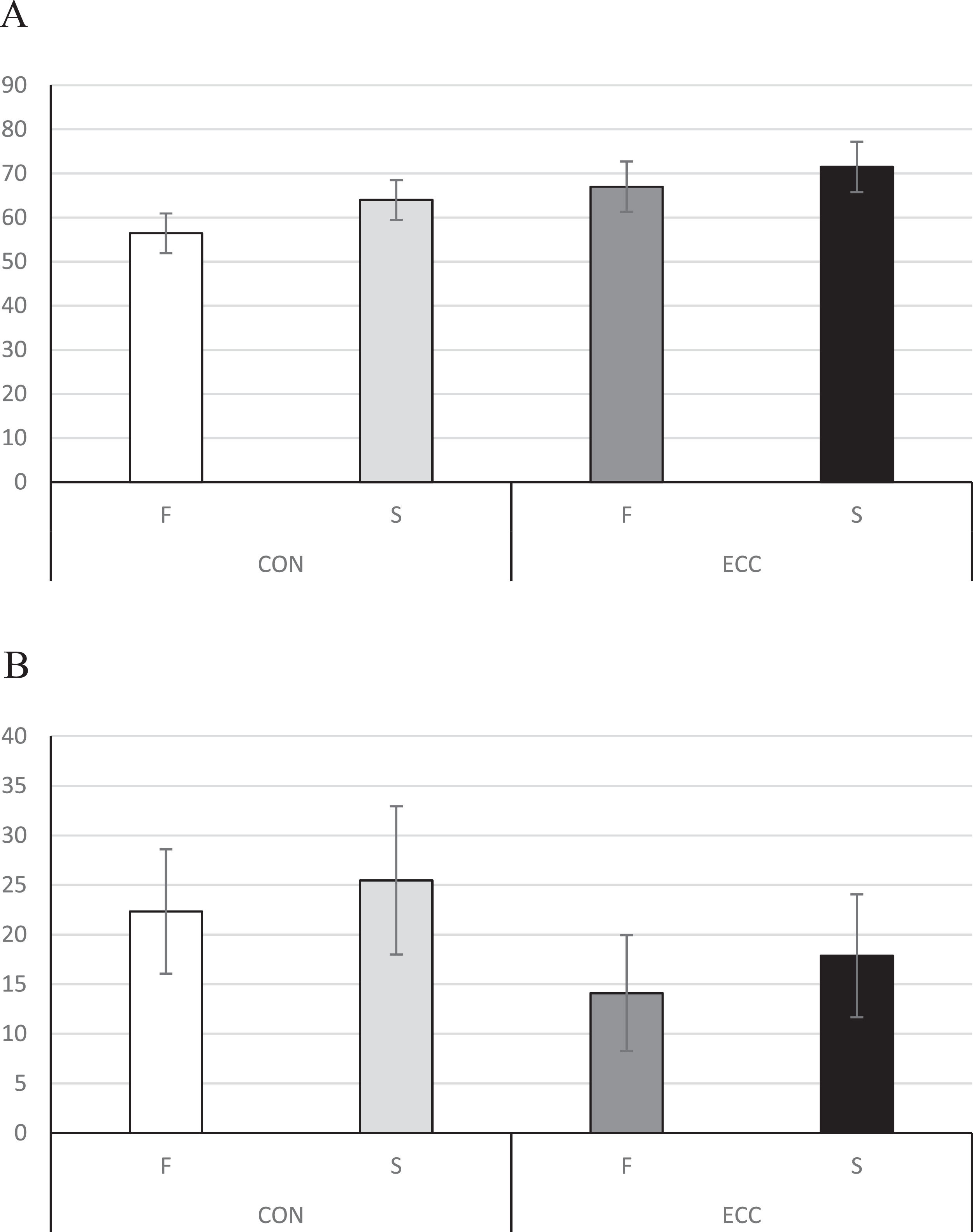
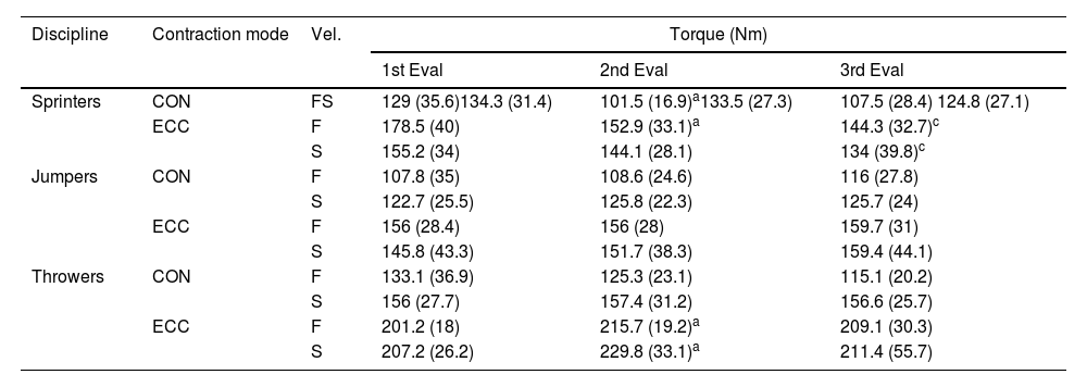
For the knee extensors, angle of PT ranged from 56 to 71º depending on the assessment speed and contraction type, while angle of PT was between 14 and 25º for the knee flexors. No statistically significant differences were found for the angle of PT between the different time points nor disciplines, the mean of the three measurements is listed for each muscle, contraction type, and velocity in Figure 1.
Functional ratios and flexors (B). Data is presented as mean ± standard deviation, with 0º indicating complete knee extension.")
Despite the variations that were observed in PT throughout the season, there were no significant differences between the three time-point measures for each calculated ratio (Table S1). Comparisons between fast and slow EXT ratios showed significant differences in favour of the slow assessment, where QF generated larger forces than HAMS (p < 0.05).
DiscussionWe prospectively investigated knee flexors and extensors peak torque, the angle at which peak torque was achieved, and functional ratios in a population of male track and field athletes throughout an entire season. The main findings were: (1) knee flexor and extensor peak torque varied significantly along the time-point measurements with moderate to large effect sizes observed (ES (d) = 0.49–0.93) (Table 2), irrespective of the discipline; (2) changes in the angle of peak torque were non-significant throughout the study. To the best of our knowledge, this is the first longitudinal study on this subject.
We observed that CON knee flexor PTs ranged from 1.5 to 1.7 Nm/kg, depending on the discipline. Our participants were slightly weaker than professional footballers in terms of CON torque production, generating 2 Nm/kg during knee flexor testing.17 Regarding knee extensor torque (Nm), our results (180.7 vs. 405.8 Nm across the disciplines) were similar to those of a recent review, where torque productions of 225 Nm at slow speed were reported.18 In another study, elite basketball players produced 147 Nm at CON 300º/s, which was again similar to our findings (127 vs.150 Nm).19
When an athlete is deemed to be ready to return to their sport, certain criteria must be satisfied to ensure they are safe and at low risk of further damage. This is especially critical after muscle injuries since these tend to be more severe and require longer periods of rehabilitation.20 Tissue healing, flexibility, the absence of pain, and functional performance must be taken into account before making the final decision.20 In addition to the aforementioned criteria, HAMS and QF PTs have to be assessed when these muscles have been injured.14
The sprinters in the present study were stronger than footballers who had suffered an HSI (CON 300º/S: 1.7 Nm/kg vs. 1.1 Nm/kg; ECC 300º/s: 2.3 Nm/kg vs. 1.8 Nm/kg).21 Similarly, footballers who had recently suffered an anterior cruciate ligament tear (ACL) displayed QF deficits in injured (CON 60º/s: 1.6 Nm/kg) and healthy (CON 60º/s: 2.3 Nm/kg) limbs before ACL reconstruction, compared with our healthy athletes (CON 60º/s: 2.5 vs. 2.9 Nm/kg). This may be due to arthrogenic muscle inhibition, where changes to efferent function contribute to inhibition of the QF motor neuron pool, limiting the capacity to generate force.22 The present study complements the literature by pointing out how disciplines differ with other sports or athletic disciplines, velocity disciplines require that force must be applied fast, therefore rate of force development might be higher in these sports whereas throwing might be more biased towards maximal strength. Therefore, when making clinical decisions regarding return to sport after QF or HAMS injury in athletics, data here presented could serve as reference to know whether an athlete is ready to train or compete again.
The angle at which HAMS PT (knee flexion 20º vs. 30º; see Angle of peak torque section) was achieved remained stable throughout the season, these results should be treated with caution since they have clinical relevance in the presence of a history of HSI and should be taken into consideration when planning a return to sport after a thigh injury. Mikami et al.21 recently observed that those athletes with a previous HSI generated significantly less eccentric torque at the last degrees of knee flexion when compared with their equivalent healthy limb. Normalised PT at 20º of knee flexion was 1.7 and 1.9 Nm/kg for the injured and healthy limbs, respectively. Athletes with a previous HSI displayed larger absolute torque deficits than uninjured pairs after an experimental fatigue-induced test.23 Similarly, those with a previous HSI generated 30% less rate of torque development during eccentric contractions when compared with a control group and uninjured limbs.24 These findings have implications for subsequent injury risk, since a partial restoration of normal function may predispose the muscle tissue to further strain or damage,25,26 particularly given that HSIs occur primarily when the knee is at almost complete extension and the muscle is fatigued.27,28
Certain limitations in the present study must be mentioned. Only adult male athletes participated; certain differences might be observed between genders or adolescent athletes, caution should be taken in generalising these findings to both young people and females. Another limitation is the usage of isokinetic dynamometry, which is not accessible in every sporting environment due to its high cost. Recently, more accessible alternatives have appeared. For instance, the Nordboard, which makes testing faster, has shown promising results.29 Limitations aside, the present study (to the best of our knowledge) is the first to record thigh muscle strength data prospectively in track and field athletes. Also, the peak torque data, which was analysed in terms of the angle at which the peak torque occurred, might assist in return-to-sport testing after an HSI. Such information is not always taken into consideration in studies of a similar nature.21,25
ConclusionAdult semi-professional male track and field athletes exhibited variations in thigh muscle peak torque throughout the season. However, the angle at which PT was achieved and functional ratios seemed to remain constant. Our observations could be used as normative data for track and field athletes both in comparison with healthy people from a performance perspective or with previously injured athletes, thus aiding in their decisions about when to return to sport.
Ethical considerationsThe local institutional review board approved this study (code: 2021.543) and all participants signed a written informed consent.
FundingThe authors report no funding.
The authors would like to thank all the participating athletes.

