


the muscular response on the main muscles of the propulsive phase of the upper limb in the crawl swimming style has been evaluated by means of tensiomyography (TMG), which was carried out through three parameters: activation time (Td), contraction time (Tc) and maximum deformation (Dm).
The aim of this study is to evaluate the hypothesis of changes produced between lactic resistance training with passive rest and lactic resistance training with active rest.
Material and methodsexperimental study with ninety swimmers in the crawl stroke, with a mean age of 20 years. The session with passive recovery and the session with active recovery presented results with significant changes after the development of each of them in the Tc in the three muscles analysed, being in the Pectoralis Major (PM), p = 0.001, in the Latissimus Dorsi (LD), p = 0.01 and in the Tríceps Brachii (TB), p = 0.047.
ConclusionAccording to the results it can be deduced that lactic resistance training with passive rest and with active rest do not influence the muscle response if we compare both trainings. Only one of the Tc values changes, while the latissimus dorsi muscle also shows changes in Td.
The great importance of lactic anaerobic work in swimming, especially for swimmers who are specialists in 50 m and 100 m events, is related to the muscular predisposition in relation to their need for energy throughout the event and training.1,2 For this reason, the lactic anaerobic training system is considered to be of vital importance when focusing on it in planning.3 Within the lactic anaerobic system, various training methods can be found which can be carried out by series with active or passive rest between them, with some authors such as Romero4 highlighting that in high-intensity work passive rest increases performance and others such as Micklewright5 indicating that this type of recovery does not affect the increase in lactate as long as this interval is no longer than 5 min, but for other authors such as Navarro-Valdivielso et al.,3 active recovery is more beneficial for the organism in relation to physiological aspects.
To analyse the muscular response of these muscles and how they respond to a session, TMG is used, which is a tool that has been used in sports with similar movement patterns such as lifeguarding6 to analyse the contractile characteristics of the superficial muscles1; This tool has been used in the processes of analysis of muscle response in other different sports, but with movement patterns similar to the one studied on this occasion, so it appears to be a useful tool capable of responding to the proposed objective; its non-invasive evaluation technique allows the mechanical response of specific muscle structures to be analysed1, and also, possible alterations in muscle response can be assessed to indicate the potential appearance of a specific type of fatigue.
The aim of this study is to compare the effect of two different types of recovery, active vs. passive, on the muscular response of swimmers in lactic resistance training on the pectoralis major, latissimus dorsi and triceps brachii.
Material and methodsThis research has been carried out in accordance with the ethical standards of good clinical practice (Declaration of Helsinki, Edinburgh revision, 2000) and respecting the aspects established in the current legislation on clinical research in relation to the convention for the protection of Human Rights.
This project has been approved by the CEIC of the Community of Madrid. It complies with Organic Law 3/2018, of 5 December, on Personal Data Protection (LOPD) and the guarantee of digital rights, which provides for the protection of personal data in the scientific research and medical trials sector. In addition, the trial has been registered at www.clinicaltrial.gov under the reference number NCT06155526.
The participants have signed a consent for the conduct of this study, for the provision of their data and also for a possible publication of their data.
ParticipantsThe sample consisted of 90 participants and was selected using convenience sampling; the chosen swimmers, male, from the community of Madrid, belonged to the junior-absolute category and the predominant stroke was the crawl.
All the participants signed the informed consent to undergo the research and also agreed to provide their data for possible publication.
The variables measured were TMG parameters such as activation time (Td), contraction time (Tc) and maximum deformation (Dm), where Td and Tc are measured in milliseconds (ms) and Dm in millimetres (mm).
Participation in this study was established according to the following inclusion criteria: being between 18 and 25 years of age, having a minimum mark or having participated by ranking in a crawl event in the territorial championship of Madrid in the current season in which this study was carried out and belonging to the male gender.
The exclusion criteria were as follows: subjects who did not meet the minimum age and did not obtain the minimum mark and did not sign the informed consent form.
Materials and instrumentsThe sessions were carried out in a 25.03 m indoor swimming pool with a minimum depth of 1.69 m and a maximum depth of 2.28 m.
For the assessment of muscle response, a tensiomiograph model TMG system 100, manufactured in Slovenia, was used. The Td, Tc and Dm variables were analysed according to the protocol proposed by Rodríguez-Matoso1 as well as the manufacturer's indications for the placement of the electrodes and the sensor with an electrical impulse below the maximum threshold stimulus.7
In relation to the stimulus used by means of the TMG, in our study a stimulus of 1 ms with an intensity of 100 m was given. The tensiomyograph can produce a current stimulation ranging from 0 to 10 mA, and the sensor has a maximum measurement length of 32 mm and a maximum velocity of 1 m/s.
ProcedureFirstly, swimmers who could a priori meet the inclusion criteria were contacted and informed of the aim of the study and given a consent form.
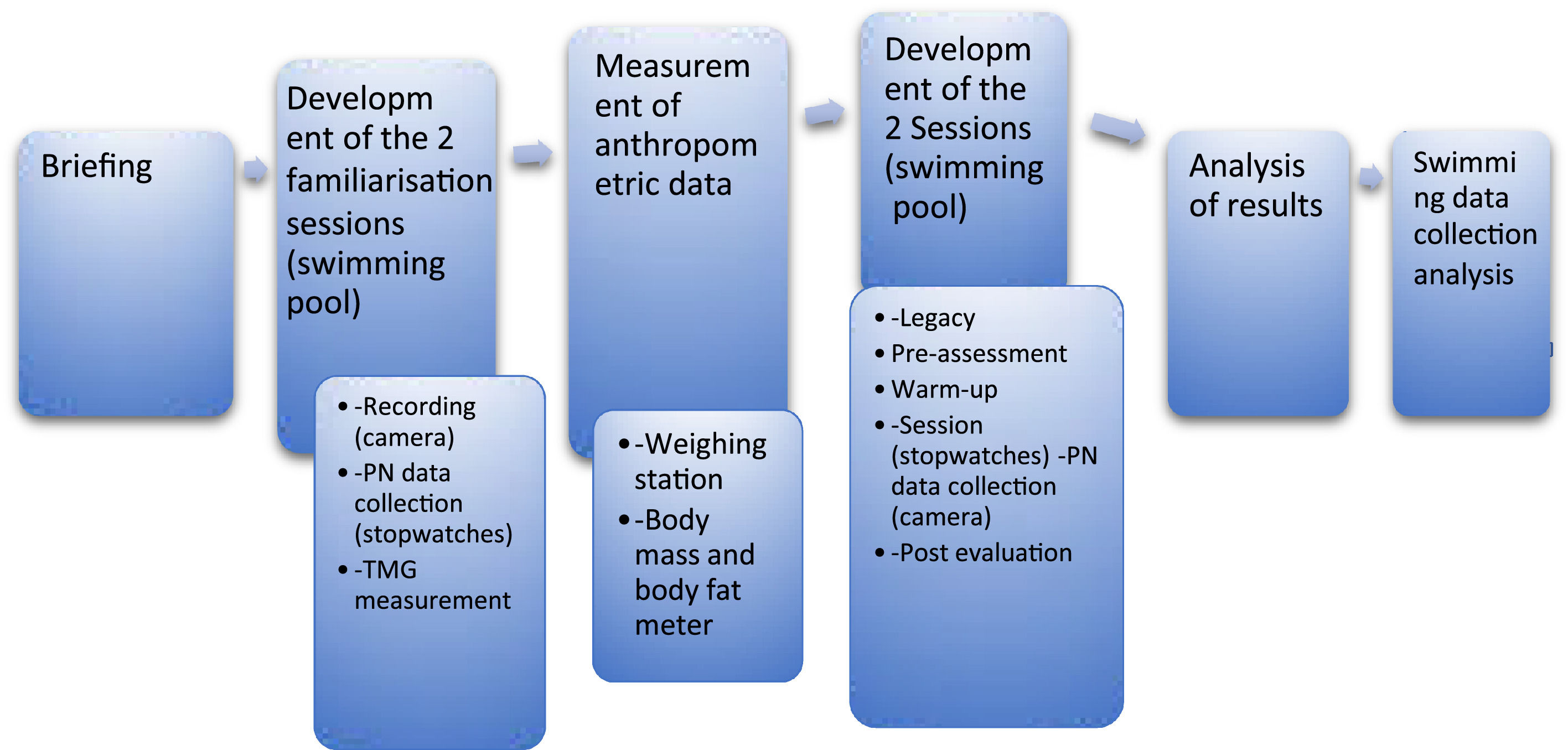
Two weeks after the meeting, two familiarisation sessions were held, in which the session with active recovery (SA) and the session with passive recovery (SP) were carried out as a test, and then repeated in the research study itself (Figure 1).

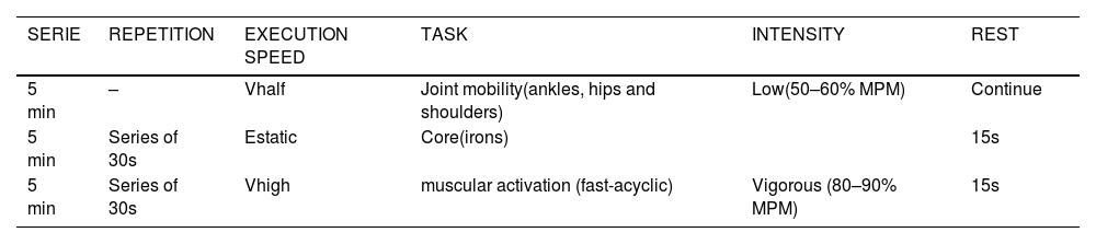
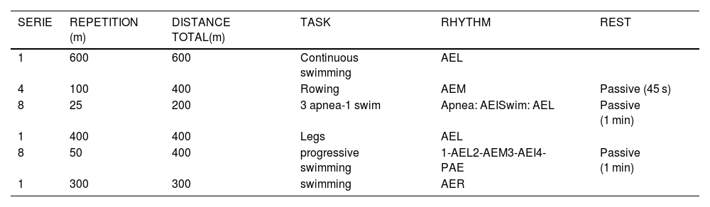
In the following fifteen days, we began with the development of each of the two lactic resistance sessions (table 3) with a dry warm-up (Table 1) and a warm-up in water (table 2) common to both sessions in which the mechanical muscular response of the PM, the DA and the TB were measured, always on the right side by means of TMG before (pre) and after (post). To ensure homogeneity in the TMG measurement times between swimmers, the athletes were previously divided into groups of 5; each group arrived with a difference of 25 min, which made it possible to analyze the mechanical response of the three muscles before training for all the subjects and, in this way, to ensure that the waiting times between the TMG measurements and the session were more homogeneous.
Warming in water.
Note: s (seconds). min (minutes). m (metres). AER (aerobic recovery). AEL (aerobic light). AEM (medium aerobic). AEI (intense aerobic). PAE (aerobic power).
Description of the main part of each session.
| MAIN PART OF EACH SESSION | |||
|---|---|---|---|
| Session SA | 4X(4 × 25 m) /5 s/4min | Work: 95–100% MMP 100Rest: AER | Active rest series |
| Session SP | 4X(4 × 25 m) /5 s/4min | Work: 95–100% MMP 100 | Passive rest series |
Note: s (seconds). min (minutes). m (metres). SA (set session with active rest). SP (set session with passive rest). MMP 100 (100 m personal best). AER (aerobic recovery).
Once the corresponding group had arrived at the pool, the pre-analysis was carried out and then the warm-up was started with the warm-up out of the water and the corresponding session in the water (Table 1), the development of the main work, which was carried out in crawl, the swimmer in the first repetition of each series left the starting platform, and then continued the training by carrying out the rest of the work from the water. The rhythms of each repetition were recorded with three timekeepers to assess the homogeneity of the swimming rhythms of each series.
In the session with active rest, the swimmer carried out the rest with a series of continuous swimming at a regenerative intensity and aerobic recovery (AER), in the event that the regenerative swimming time was completed and the swimmer was not at the starting point of the next series, he/she was allowed to continue until reaching that point. On the other hand, in the session with passive rest, the athlete remained seated out of the water and in a static manner without making any effort between each series.
Then, once the session was over, the mechanical response of the analyzed muscle was analyzed again before returning to the water to calm down.
AnalysisSeveral analyses were carried out using the statistical software IBM SPSS Statistics V.29.0.0.0 (IBM SPSS, Chicago, IL).
The descriptive statistics used were: mean, standard deviation, maximum and minimum values and 95% confidence intervals.
Multivariate repeated measures factor analysis was used for the inferential analysis. To obtain the results of the mechanical characteristics of the muscles analyzed, 2 times factors (Pre, Post) and 2 training factors (SA, SP) were used. In addition, a pre-post analysis was carried out on the Td, Tc and Dm parameters of the three muscles analyzed in relation to 2 factors, time and time by training.
The effect size was calculated using the eta partial square test, establishing small (<0.06), medium (0.06–0.13), and large (>0.13) effect sizes (8).
A p-value of 0.05 was set as the level of significance. After checking the normality of the sample using the Shapiro-Wilks test, the variables that did not follow a normal distribution model were analyzed using Q-Q plots and histograms to rule out the presence of extreme values in the distribution.8
ResultsThe sample characteristics are reflected in the descriptive statistics shown in Table 4.
General descriptive statistics of the sample.
Note: (mean value of each variable). SD (standard deviation). CI (confidence interval). Kg (kilograms). cm (centimetres). BMI: Body Mass Index s (seconds).
Pectoralis major:
In relation to the multivariate analysis of the PM (table 5), a significant change has been observed in the time factor with respect to the mechanical characteristics of the PM with a large effect size, being the value of F (3,27) = p < 0.001; =0.994; 1-ß=1. In table 6 we can observe the univariate analysis of the pectoralis major in relation to time and time-training.
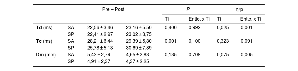
Univariate analysis of the pectoralis major in relation to time and time-training.
Note: Tc (contraction time). Td (deformation time). Dm (maximum deformation). Ti (time). Ennto. x Ti (time training). p (significance). η²p (partial eta squared).
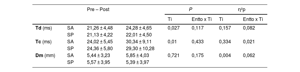
Regarding the multivariate analysis (table 7), a significant change was observed in the time factor with respect to the mechanical characteristics of the DA with a large effect size, being F (3,27) = p < 0,001; =0,990; 1-ß=1).
In relation to the time*training interaction, no significant differences in the mechanical characteristics have been observed. In table 8 we can see the univariate analysis of the latissimus dorsi, as well as the descriptive values.
Univariate analysis of latissimus dorsi in relation to time and training time.
Note: Tc (contraction time). Td (deformation time). Dm (maximum deformation). Ti (time). Ennto. x Ti (time training). p (significance). η²p (partial eta squared).
Analysis of the triceps brachii:
Regarding the multivariate analysis (table 9), a significant change in the time factor has been observed regarding the mechanical characteristics of the TB with a large effect size, being F (3,27)= p < 0,001; =0,996; 1-ß=1).
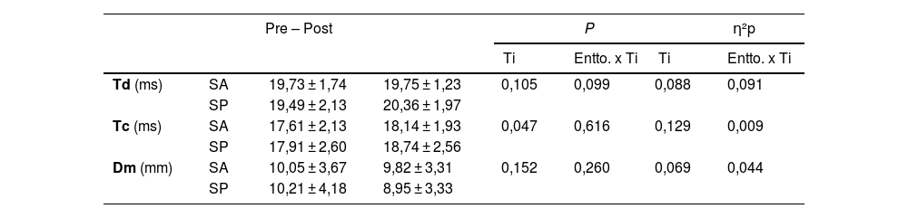
In relation to the interaction of time*training no significant differences in the mechanical characteristics have been observed. In Table 10 we can see the univariate analysis of the triceps brachii, as well as the descriptive values with significant differences in the Tc in the pre and post-training (p = 0.047) and in the rest of the values there were no significant differences.
Pairwise analysis of triceps brachii in relation to time and training time.
Note: Tc (contraction time). Td (deformation time). Dm (maximum deformation). Ti (time). Ennto. x Ti (time training). p (significance). η²p (partial eta squared).
It should also be noted that the time evaluation refers to the pre and post in one session and the time*training refers to the comparison of the pre between both trainings and the post between both trainings.
DiscussionThe PM is considered to be a muscle of great influence in the propulsive phase of the crawl stroke swim.9 Accordingly, this muscle should present changes in its mechanical response as proposed by several authors,10,11 these changes in Tc can be modified according to the type of active fibers, the state of fatigue or muscle activation. In this case, it may be due to a deformation in the execution of the technique, which would cause an increase in resistance leading to a muscular imbalance with a subsequent development of possible muscular injuries.10
Monteiro et al.11 found that the MP showed changes in its mechanical response after maximal strength training, in this case, Tc decreased significantly and Dm increased, which did not indicate a good response to strength training.
In the interaction of the time factor with the training, we found no significant differences in any of the parameters, which indicates that there is no change based on the sessions carried out,12 however, significant differences were found in reference to the training time factor, although this may be due to the different methodology with regard to what was carried out in our study.
After the completion of each of the two sessions, according to the significant differences obtained from the TMG parameters, we can deduce that this muscle is in a state of rigidity presenting signs of acute muscle fatigue according to the nomenclature of Farto (2003),13 this may be because, as indicated by Figueiredo et al.14 in a study using EMG and in which they state that the PM is a muscle with a high incidence in this style and is involved in the inward movement phase in the propulsive phase of the stroke, so this added to the load received in each of the sessions, could be the causes that motivate this change in muscle mechanics.
The DA is also a muscle of great involvement during the inward movement phase of the underwater phase of the crawl stroke14; which we can relate to the significant differences found in this study in relation to the possible presence of muscle fatigue with respect to the time factor. A study by Lomax et al.15 states that fatigue in the inspiratory muscles can affect the activity of DA work, which could lead us to the conclusion as to why we found differences in our study between pre and post. On the other hand, a study by Stirn et al.16 (2011) concludes that in crawl swimming, the dragging of the resistance offered induced the presence of fatigue in the DA.
Coinciding with the results obtained in our study after the two sessions, Costill et al.17 suggest in their study that in a training programme, although the swimmers experienced muscle fatigue, this did not vary according to the training programme developed.
About the results observed in our research, we can see that there is a significant difference in the Tc, so we can conclude that this difference indicates a greater activation of type I fibres.18
Concerning the TB, in the last propulsive phase of the crawl stroke, before starting the recovery, the TB acquires great importance,14 which may lead us to deduce that due to this, we observed a significant difference in the time factor after the session in relation to the mechanical characteristics of the muscle.
Ganter et al.,19 for their part, indicate that muscle fatigue in swimmers is present in high-load training sessions, coinciding with the results obtained in this study. Strength work influences the contractile capacity and stiffness of this musculature,9 although in this case surfers have been evaluated, so the biomechanical pattern of this muscle may interfere in a different way to that of swimmers.
On the other hand, with the analysis of each parameter of the TMG, we can see how the Tc, depending on the training session carried out, behaves differently, presenting a significant difference between the values obtained. In principle, the intervention of this muscle in the final phase of the pull of the stroke generates acute muscle fatigue, which appears after a training session in which it exceeds the level of tolerance to effort in the muscle, reducing the temporary work capacity.20
Hormigo21 also suggests in his study on judo players in relation to maximum strength and power work that the work of the TB in the Tc parameters underwent modifications after the sessions, which is similar to the results obtained in our work. In this case, the movement pattern also differs from that of the TB in the crawl style.
Moving on to the analysis of the muscular state after the execution of each session, we can see that in the SA and SP, the muscle presents a state of rigidity with acute muscular fatigue.
The TB, as mentioned above, has a great influence on muscle fatigue in the crawl style14; a study by Conceicao et al.22 concluded in the analysis by means of EMG of a group of swimmers in a 200 m crawl race and in which the TB presented fatigue throughout the race, which suggests that this would be in agreement with our results, clarifying that the volume worked in this study was different to ours.
ConclusionIn relation to the comparison of training with passive rest vs. training with active rest, it should be noted that the three muscles analyzed present changes in muscular response after each session, these changes occur in one of the Tc values, while the latissimus dorsi muscle also presents changes in Td.
This research did not receive any specific grant from funding agencies in the public, commercial, or not-for-profit sectors.
All authors should have made substantial contributions to all of the following: the conception and design of the study, acquisition of data, analysis and interpretation of data, drafting the article or revising it critically for important intellectual content and final approval of the version to be submitted.

